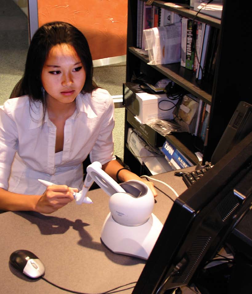
above: For Ohio State medical student Dinah Wan, the force-feedback device creates the pressure and resistance experienced during surgery. Without a virtual simulation, medical residents would learn surgery techniques by working on cadaveric specimens and through apprenticeships in an operating room.
inset: The skull’s temporal bone, as viewed here in simulation, encases some of the body’s tiniest and most delicate sensory structures.
The next generation of surgeons — many who grew up playing video games — are using real-time, interactive computer simulations to learn delicate surgical techniques required for operations on the human skull.
Called the Virtual Temporal Bone Project, the system was developed as a collaborative project between physicians and researchers at Nationwide Children’s Hospital, The Ohio State University Department of Otolaryngology and the Ohio Supercomputer Center.
Without virtual simulation, medical residents would learn surgery techniques by only working on a few cadaveric specimens and through apprenticeships in an operating room.
The complexity of the skull’s temporal bone makes surgery exceptionally challenging to master. The bone forms a major portion of the skull base and encases some of the body’s most minute and sophisticated sensory structures, including the cochlea and semicircular canals. Meanwhile, it sits intimately beside the carotid artery, jugular vein, cerebral cortex, brainstem and cranial nerves. Surgeons drill into the temporal bone to treat ailments such hearing loss, vertigo, chronic ear infections or tumors.
“The virtual bone simulation is a fantastic teaching tool,” said Dr. Laura Matrka, a second-year resident at The Ohio State University Medical Center.
“I can use the tutorial mode to see the layers and structures under the bone. Then I can switch to a practice session and drill on the structure for a very lifelike experience.” Co-investigators Dr. Gregory Wiet, a pediatric otolaryngologist, head and neck surgeon with Nationwide Children’s Hospital and an associate professor of otolaryngology and biomedical informatics at The Ohio State University, and Don Stredney, director of the Interface Lab and a research scientist in biomedical applications at the Ohio Supercomputer Center, have begun a national multi-institutional validation study to determine how medical students trained with the simulator compare to those trained using traditional methodology.
“We want to know for certain that we’ve created a safer, more effective way to learn fundamental techniques,” Dr. Wiet said.
For Dr. Matrka, the system has met its mark. “In terms of learning the anatomy and organizing in my mind the way I want to go about doing a temporal lobe dissection, it’s excellent,” she said.
--
Project leads:
Gregory Wiet, M.D., Nationwide Children’s Hospital & The Ohio State University, & Don Stredney, OSC
Research title: Validation/dissemination of virtual temporal bone dissection
Funding source: National Institute on Deafness & Other Communication Disorders, National Institutes of Health, 1-R01-DC06458-01A1